

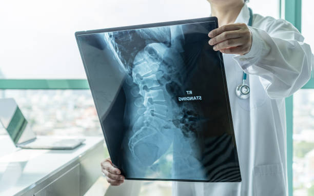
Why do bones fail?
Osteoporosis is a widespread disease. Every third woman and every fifth man are affected by bone loss with advancing age. A frequent consequence of this is a fracture of the femoral neck – a painful injury that massively impairs the quality of life of those affected. Patients must reckon with longterm loss of mobility. Long bed rest and the associated often poor general condition even lead to an increased mortality rate.
The disease causes a loss of bone mass due to an imbalance in the natural remodelling process in the tissue and changes in bone quality. These changes affect the microstructure, density of microcracks and tissue properties.
Bones have an extremely complex structure. If, for example, a thigh bone is sawed open, it can be seen that it consists of a hard outer layer and a porous filling. Under the microscope, cylindrical structures of concentric lamellae are visible inside the hard shell, arranged around central blood vessels. These individual lamellae are only a few thousandths of a millimetre thick and consist of a type of natural fibre composite material: collagen fibres in which mineral particles are embedded, embedded in a protein-containing mineral matrix. The higher the mineralization, the stiffer and more fragile the bone. This hierarchical structure allows the bones to be robust and resistant despite their relatively low density. When bones fracture, it is therefore not sufficient to consider only the density and structure of the bone at the macro level – mechanisms in all scale ranges are responsible for the fracture.
Material analysis for bone
A research group at Empa in Thun led by Jakob Schwiedrzik aims to gain a better understanding of bone failure at the lamella level. “If one only considers bone density, as is usually the case in clinical practice today, the risk of fracture for patients can be predicted relatively well on average. In individual cases, however, the results may differ considerably and the effective fracture risk may be incorrectly assessed,” explains Schwiedrzik. “We hope that our research will enable us to make more accurate predictions for each individual patient in the future”.
The researchers are using methods that are actually at home in materials research: They subject even the smallest samples of bone material containing only a single lamella to tensile and com-pression tests. They are investigating how the material fails and how the measured properties are related to the underlying microstructure. In microstructure analysis, Raman spectroscopy and transmission electron microscopes are used – highly complex instruments that make it possible to precisely observe structural changes in the test objects.
“At the moment, the production and testing of a single bone sample still requires a great deal of time – especially for tensile tests,” explains Schwiedrzik. To do this, samples with a defined geometry must first be produced from the material used using a focused ion beam. In order to be able to analyze more samples in less time in the future and to enable statistical evaluation of the experiments, a large part of the current work consists of automating the sample heart position and developing our own measurement setups.
Personal diagnosis
The question of how the methods developed can be used for clinical studies is exciting. A project is currently underway involving researchers from the Inselspital Bern, the University of Bern, ETH Zurich and Empa. Bone material from patients who have received a hip implant is being investigated. This material will be analysed on several length scales. The aim is to collect data on micromechanical properties, microstructure, cell activity and metabolism and to correlate these with clinical findings and patient data using machine learning. The resulting database will make it possible to quantify the bone quality of a patient and include it in the diagnosis.
Source: Empa
Full bibliographic information
Mechanical properties of cortical bone and their relationships with age, gender, composition and microindentation properties in the elderly
Bone, Volume 93, December 2016, Pages 196-211
https://doi.org/10.1016/j.bone.2015.11.018
