
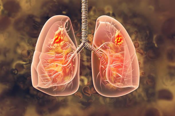
Researchers identify new biomarkers of the most frequent lung cancer
Researchers of the University of Barcelona and the Institute of Bioengineering of Catalonia (IBEC) have identified new biomarkers for non-small cell lung cancer, the most common lung cancer. The results, published in the journal Modern Pathology, have been obtained thanks to a new technique that allows quantitative analysis of patient samples. It is a pioneering methodology that is cheaper and easier to apply than the traditional ones in hospitals and other clinical settings.
The study notes that certain features of the collagen fibers —one of the most abundant components regarding cancer cells— would be a potential indicator for the diagnosis and prediction of the evolution of the disease.
“Our new tool can improve the clinical management of surgical patients with this type of cancer, since it identifies those at an increased risk of relapse and, therefore, can benefit from a comprehensive monitoring and even neoadjuvant therapies (complementary to the main treatment)”, notes Jordi Alcaraz, lecturer at the Faculty of Medicine and Health Sciences of the UB and researcher at IBEC, who led the study together with Joan Montero, researcher at the same faculty, and Josep Samitier, professor at the Faculty of Physics and director of IBEC.
The study includes the participation of researchers from Hospital Clínic de Barcelona, the Respirtory Diseases Networking Biomedical Research Centre (CIBERES), the Bioengineering, Biomaterials and Nanomedicine Networking Biomedical Research Centre (CIBER-BBN), the University Hospital 12 de Octubre (Madrid) and the University Hospital Parc Taulí (Sabadell).
What is the role of collagen fibers in tumor progression?
Lung cancer is the main cause of death related to cancer in both men and women worldwide, with a five-year survival rate of 18%. Most of these patients are diagnosed with non-small cell lung cancer, which is subdivided into adenocarcinoma, squamous cell carcinoma and other less common subtypes. There is increasing evidence of the essential role of the collagen fiber-rich environment surrounding cancer cells in the progression of these cancers and other solid tumors.
In this tumor context, high expression of type I collagen has been associated with poor prognosis and increased risk of metastasis. “This evidence has pointed to fibrillar collagens as an important potential source of cancer-relevant biomarkers and has sparked therapeutic interest in understanding their roles in tumor development”, says Jordi Alcaraz.
Faced with this challenge, researchers have developed and validated a new digital pathology approach — that is, the study of disease with digital tools — to quantitatively analyze collagen fibers in tissue samples from patients with non-small cell lung cancer.
A more accessible technique for hospital pathology units
The new methodology is based on digitized images of patient biopsies stained with a dye called picrosirius red (PSR) and imaged with polarized light. The researchers use the open-source software CT-FIRE to automatically segment the individual fibers in the images to quantify relevant characteristics such as length, width or straightness.
“The standard methodology for analysing collagen fibers is based on an advanced microscopy technique called second harmonic generation, which requires a double-photon confocal microscope, a type of microscope which is expensive and needs an expert operator”, he notes. “In contrast — he continues —, our tool is a cheaper and easier-to-incorporate approach for pathology units, since it uses an inexpensive stain (PSR), free software such as CT-FIRE and a microscopy technique accessible to most hospitals, such as polarized light”.
Collagen density, an indicator of poor prognosis
The researchers have applied this methodology to analyze 195 samples from patients with adenocarcinoma and squamous cell carcinoma. The results show that fiber straightness would be a potential biomarker for diagnosing the disease while density would be a poor prognostic indicator. Moreover, the prognostic value of collagen density would be “independent of the clinical stage of the tumor, and it shows that analyzing collagen fibers provides additional relevant information”, stresses Jordi Alcaraz.
Abnormal stiffening of tissues in patients with adenocarcinoma
The new methodology has also made it possible to compare the characteristics of collagen fibers in tissue samples from patients with non-cancerous tissue samples. Thus, for the first time, they have been able to describe quantitatively the changes that take place in the organization of collagen in non-small cell lung cancer.
The results show that, in tumor tissue samples, there is an increase in straightness, length and width that indicates an abnormal stiffening of the tissue, especially in patients with adenocarcinoma. According to the researchers, this stiffening would be related to different mechanisms to avoid the immune system response and, consequently, to tumor progression in this type of cancer. “This opens the door to identifying new therapies directed against abnormal stiffening”, concludes the researcher.
This study was funded by the State Research Agency of the Spanish Ministry of Science and Innovation, La Caixa and Cellex Foundation.
https://linkinghub.elsevier.com/retrieve/pii/S0893395223000601
Full bibliographic information
Published on 20/06/2023 by Universidad de Barcelona
Authors: Almici, E.; Arshakyan, M.; Carrasco, J. L.; Martínez, A.; Ramírez, J.; Enguita, A. B.; Monsó, E.; Montero, J.; Samitier, J.; Alcaraz, J. About: «Quantitative Image Analysis of Fibrillar Collagens Reveals Novel Diagnostic and Prognostic Biomarkers and Histotype-dependent Aberrant Mechanobiology in Lung Cancer», Modern Pathology, march 2023.
DOI: https://doi.org/10.1016/j.modpat.2023.100155


